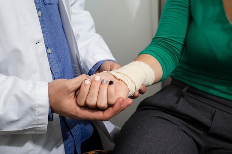
Doctor treating a person with a stress fracture.
A stress fracture is not the kind of injury most people picture when they hear the word fracture. There is no single dramatic fall or collision. No obvious moment of injury. Instead, a stress fracture develops silently, the result of repetitive force on bone over a period of time, with pain that begins as a mild ache and gradually becomes impossible to ignore.
Stress fractures are common among athletes, military recruits, and anyone who has recently increased their physical activity significantly. They are also seen in people with weakened bones due to nutritional deficiencies or medical conditions.
Left untreated, a stress fracture can develop into a complete fracture, a far more serious injury requiring significantly longer recovery. Understanding stress fractures early is essential.
This article explains what stress fractures are, how they develop, how they are diagnosed, and what treatment and recovery involve.
A stress fracture is a small crack or severe bruising within a bone caused by repetitive mechanical loading. Unlike a traumatic fracture, which results from a single high-energy impact, a stress fracture results from cumulative, repeated stress that exceeds the bone's capacity to repair itself.
Bone is a living tissue. Under normal conditions, the body constantly breaks down and rebuilds bone through a process called remodelling. When physical stress on a bone increases faster than the body can remodel and strengthen it, microscopic cracks accumulate and a stress fracture develops.
Stress fractures most commonly affect the weight-bearing bones of the lower body:
They can also occur in the spine (stress fractures of the vertebrae, called spondylolysis) and, less commonly, in the upper limbs in athletes such as throwers or rowers.
The most common cause. When the intensity, duration, or frequency of physical activity increases too rapidly, the bone does not have sufficient time to adapt and strengthen.
Classic scenarios include:
Running on concrete or other hard surfaces increases the impact force transmitted to bones with each step. Uneven surfaces also create irregular loading patterns that stress specific areas of bone more than others.
Shoes that lack adequate cushioning or support fail to absorb impact forces effectively. As a result, a greater proportion of that force is transmitted directly to the bones. Worn-out running shoes are a significant and frequently overlooked risk factor.
Abnormal foot mechanics such as flat feet (overpronation), high arches (underpronation), or leg length discrepancy, alter how forces are distributed across the bones during movement. This can concentrate stress in specific areas and increase fracture risk.
Any condition that reduces bone density or impairs bone remodelling increases susceptibility to stress fractures:
Bones displaying signs of osteomyelitis.
The characteristic feature of a stress fracture is pain that follows a predictable pattern. Learning to recognise this pattern is important for early diagnosis.
This is the hallmark symptom. In the early stages, pain is only present during activity, running, walking, or training. It eases with rest.
As the injury progresses, pain begins earlier in the activity, becomes more severe, and takes longer to resolve with rest. In late-stage stress fractures, pain may be present at rest or even at night.
There is a specific, pinpoint area of tenderness directly over the bone. Pressing on this spot reproduces the pain. This distinguishes a stress fracture from more diffuse conditions such as shin splints.
Mild swelling over the affected area is common. Bruising may or may not be visible.
For lower limb stress fractures, attempting to hop on the affected leg typically reproduces or significantly worsens the pain. This is a useful clinical screening tool.
Unlike traumatic fractures, there is no single moment of injury. Pain develops gradually over days to weeks. Many athletes initially dismiss it as normal training soreness and continue exercising, which worsens the injury.
A thorough history and physical examination, assessing the location of tenderness, the pattern of pain, and the training history, is the essential first step.
X-ray is usually the first imaging test ordered. However, it has significant limitations for stress fractures. In the early stages, and for up to two to four weeks after the fracture develops, plain X-rays are often normal. The fracture line only becomes visible once the healing process has begun and new bone formation is visible.
A normal X-ray does not rule out a stress fracture.
MRI is the gold standard for diagnosing stress fractures. It is highly sensitive and can detect bone marrow oedema (the early-stage bone stress response) before a fracture line is visible on X-ray. MRI also identifies high-risk fractures and distinguishes stress fractures from other soft tissue injuries.
A bone scan detects increased metabolic activity in bone, which is present in stress fractures. It is highly sensitive but less specific than MRI, as other conditions can also show increased activity.
Used in specific situations, particularly for high-risk sites such as the navicular or femoral neck, where detailed assessment of fracture geometry is required.
Not all stress fractures are equal. Orthopaedic surgeons classify them into low-risk and high-risk categories based on the likelihood of healing with conservative management and the risk of complete fracture.
These occur in well-vascularised bone under compressive loading and heal reliably with conservative treatment.
Examples include:
These occur in bone with poor blood supply, under tensile loading, or in locations where displacement is more likely. They have a higher rate of non-union (failure to heal) and a greater risk of progression to complete fracture.
Examples include:
High-risk stress fractures often require surgical consultation and more aggressive management.
Treatment is guided by the location, severity, and risk classification of the fracture, as well as the patient's overall health and activity goals.
This is the cornerstone of treatment for all stress fractures. The affected bone must be protected from the forces that caused it.
Surgery is recommended for high-risk stress fractures and those that have failed to heal with conservative management.
Procedures include:
The goal is to stabilise the fracture, promote healing, and allow earlier return to activity.
Bone healing requires adequate nutrition. During recovery:

Foods rich in vitamin D for bone and immune health.
Physiotherapy plays a critical role in full recovery and prevention of recurrence.
Recovery time varies considerably depending on the location, severity, and the individual's bone health.
Return to sport should only occur under medical supervision, following clinical reassessment and where appropriate, imaging confirmation of healing.
Most stress fractures are preventable with sensible training practices.
This depends on the location and severity. Some low-grade stress fractures allow limited walking with a protective boot. Others, particularly high-risk fractures such as the femoral neck or navicular, require complete non-weight-bearing. Always follow the advice of your treating doctor.
Both cause lower leg pain that worsens with activity. The key difference is localisation. Shin splints typically cause a diffuse ache along the inner border of the tibia. A stress fracture causes a very specific, pinpoint area of tenderness directly over the bone. MRI is the definitive way to distinguish the two.
A stress fracture will not heal if the loading that caused it continues. With appropriate rest and activity modification, most low-risk stress fractures heal well. Without rest, they progress to complete fractures, a much more serious injury.
Most stress fractures do not require surgery. However, high-risk fractures particularly those of the femoral neck, navicular, and fifth metatarsal, frequently require surgical intervention to ensure proper healing and reduce the risk of complete fracture.
Yes. Recurrence is common, particularly if the underlying causes, rapid training progression, poor nutrition, biomechanical issues, or inadequate footwear are not addressed. A structured rehabilitation programme and a gradual return to training significantly reduce recurrence risk.
Yes. Women, particularly those with the Female Athlete Triad, low energy availability, menstrual irregularities, and low bone density, are at significantly higher risk. Post-menopausal women with osteoporosis are also at increased risk. Hormonal factors affecting bone density play an important role.
Experiencing persistent bone pain or suspected stress fracture? Do not ignore it. Early diagnosis prevents a minor injury from becoming a major one.
Prakash Hospital, Noida is a NABH-accredited Centre of Excellence for Trauma and Orthopaedics, equipped with advanced imaging including MRI, CT Scan, and bone density (DEXA) assessment.
Our expert orthopaedic team provides accurate diagnosis and personalised treatment plans.
Call us at: +91 88260 00033
Website: www.prakashhospitals.in
Address: D-12A, 12B, Sector 33, Noida
Expert care when it matters most.
We offer expert care across key specialties, including Medicine, Cardiology, Orthopaedics, ENT, Gynaecology, and more—delivering trusted treatment under one roof.

Prakash Hospital Pvt. Ltd. is a 100 bedded NABH NABL accredited multispecialty hospital along with a center of trauma and orthopedics. We are in the service of society since 2001.
OUR SPECIALITIES
Patient Services
PROCEDURES
Contact Us
D – 12A, 12B, Sector-33, G. B. Nagar, Noida, Uttar Pradesh 201301
+91-8826000033


© 2026 All rights reserved.
Designed and Developed by Zarle Infotech