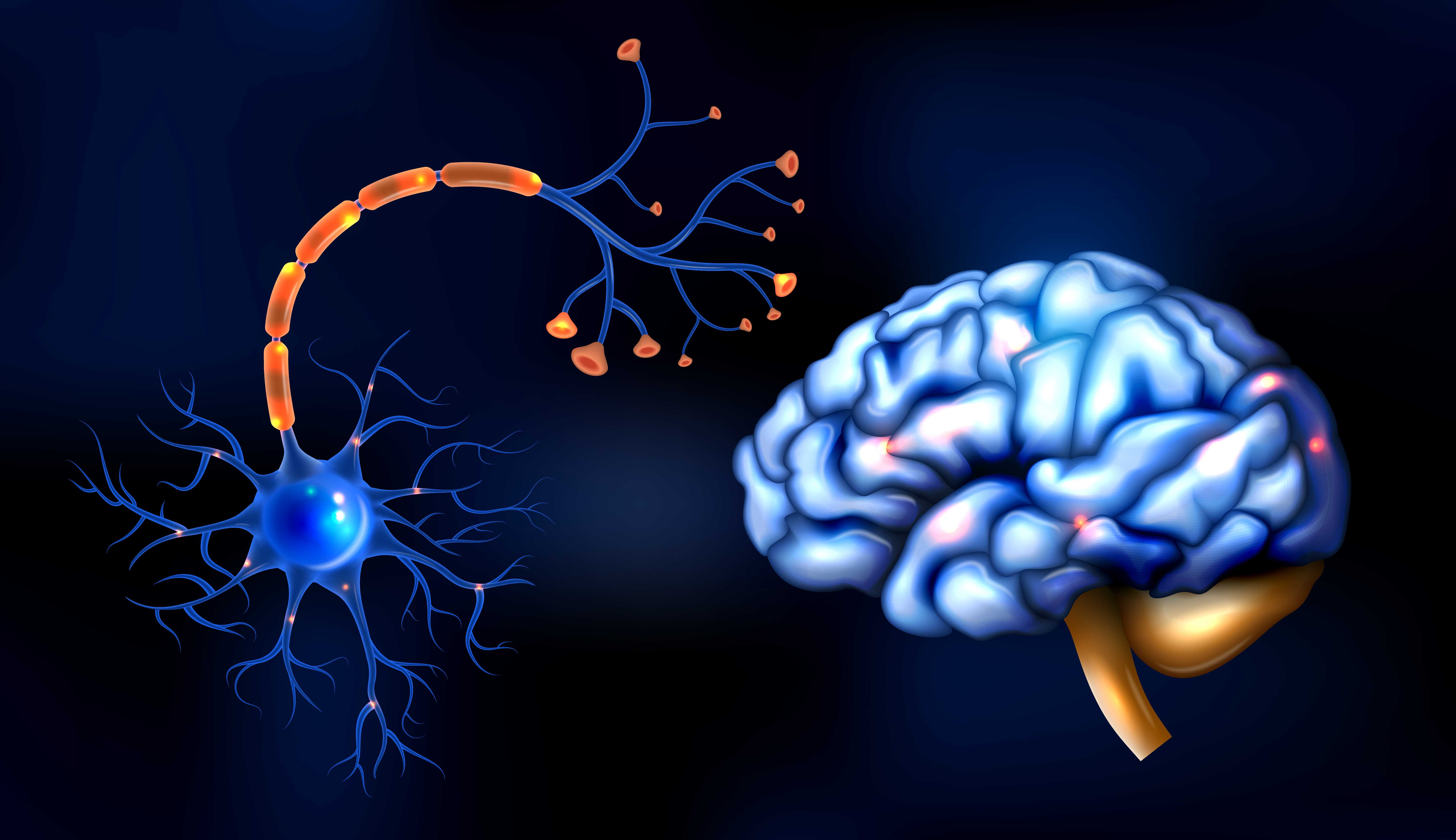
Illustration of a human brain.
A brain haemorrhage is one of the most life-threatening medical emergencies a person can experience. Every minute without treatment, approximately 1.9 million neurons are lost. The brain, unlike most organs, cannot regenerate lost cells.
Speed is everything.
Yet brain haemorrhages are frequently misidentified or ignored. The warning signs are sometimes subtle. Some people attribute the symptoms to exhaustion, a migraine, or stress and wait to seek help. That delay can be fatal or can mean the difference between full recovery and permanent disability.
This article explains what a brain haemorrhage is, the different types, the warning signs, the causes, and what emergency treatment involves.
A brain haemorrhage also called an intracranial haemorrhage, is bleeding that occurs within or around the brain. It is a type of stroke.
When a blood vessel in the brain ruptures, blood leaks into the surrounding tissue. This causes two forms of damage:
Brain haemorrhages account for approximately 10 to 15% of all strokes. They are significantly more dangerous than ischaemic strokes (those caused by a blockage), with higher rates of death and disability.
Bleeding occurs directly within the brain tissue itself. It is the most common type of haemorrhagic stroke.
Bleeding occurs in the space between the brain and the thin tissue covering it (the subarachnoid space).
Bleeding occurs between the brain and its outermost covering (the dura mater).
Bleeding occurs between the skull and the outer covering of the brain.
Brain haemorrhage warning signs vary depending on the location and type of bleeding. However, there are key symptoms that should always trigger immediate emergency response.
A haemorrhage, particularly a subarachnoid haemorrhage, classically presents with a headache that patients describe as a thunderclap: sudden in onset, reaching maximum intensity within seconds, and far more severe than any headache previously experienced.
This is often called the worst headache of my life. This description, from any patient, demands immediate emergency evaluation. It should never be attributed to migraine or tension headache without ruling out subarachnoid haemorrhage first.

A man at his desk with hands on his head, having a headache, representing brain fog, difficulty concentrating and cognitive fatigue.
One-sided weakness or numbness of the face, arm, or leg is a cardinal sign of stroke, including haemorrhagic stroke. It reflects damage to the motor cortex or its connections.
A person who was entirely well and suddenly becomes confused or difficult to rouse must be treated as a medical emergency.
Speech disturbance in combination with other neurological symptoms is a red flag for stroke.
A new-onset seizure in an adult with no history of epilepsy, particularly one followed by confusion, weakness, or headache can indicate intracranial bleeding.
A sudden collapse with loss of consciousness, particularly after a head injury, requires immediate emergency assessment.
The FAST acronym is a widely used and effective tool for identifying stroke — including brain haemorrhage.
Chronic hypertension is the single most important risk factor for intracerebral haemorrhage. It causes progressive damage to the walls of small blood vessels within the brain, making them fragile and prone to rupture. Controlling blood pressure is the most effective preventable measure against brain haemorrhage.
An aneurysm is an abnormal bulge in the wall of a blood vessel. Cerebral aneurysms often develop silently and rupture without warning, causing a subarachnoid haemorrhage. The risk of rupture increases with the size of the aneurysm, smoking, and high blood pressure.
An AVM is an abnormal tangle of blood vessels connecting arteries and veins, bypassing the normal capillary system. AVMs are usually congenital. They can rupture and cause haemorrhage at any age, including in young adults and children.
Traumatic brain injury from road accidents, falls, sports injuries, or violence is a leading cause of subdural and epidural haemorrhages. Elderly individuals are particularly vulnerable because age-related brain shrinkage stretches the bridging veins, making them more likely to tear with even minor trauma.
Anticoagulant medications such as warfarin, aspirin, clopidogrel, and newer direct oral anticoagulants, reduce the blood's ability to clot. In patients on these medications, any haemorrhage is harder to control and more likely to expand.
Conditions that impair normal clotting such as haemophilia, thrombocytopenia, or leukaemia, increase the risk of intracranial bleeding.
In elderly individuals, amyloid protein deposits in the walls of small blood vessels make them fragile and prone to rupture. This is an important cause of recurrent lobar haemorrhages in older patients.
Both primary brain tumours and metastatic deposits can bleed, causing haemorrhage within or around the tumour.
If you suspect a brain haemorrhage in yourself or someone else, call emergency services without delay. Do not drive yourself to hospital. Do not wait to see if symptoms improve. Every second matters.
Treatment depends on the type, location, and size of the haemorrhage, as well as the patient's condition on arrival.

Person undergoing a medical scan.
For many brain haemorrhages, particularly smaller intracerebral haemorrhages in stable patients, treatment is primarily medical:
Surgery is required in specific situations:
For survivors, rehabilitation begins as soon as the patient is medically stable, often within 24 to 48 hours of admission.
The goal is to maximise recovery of function lost due to the haemorrhage. This involves:
Recovery after brain haemorrhage is highly variable. Some patients recover with minimal deficits. Others face significant, permanent disability. The extent of recovery depends on the size and location of the haemorrhage, the patient's age, and the speed with which treatment was received.
Many brain haemorrhages are preventable. The following measures significantly reduce risk:
A Brain Haemorrhage Is a Medical Emergency. Every Second Counts. Prakash Hospital, Noida provides 24-hour emergency care with advanced diagnostic imaging including CT Scan and MRI, critical care facilities, and specialist medical teams equipped to manage neurological emergencies.
If you or someone you know is showing warning signs, act immediately.
Call emergency services first. Then call us at +91 88260 00033 or visit www.prakashhospitals.in Located at D-12A, 12B, Sector 33, Noida.
Fast action saves lives and protects the brain.
Yes. Many people survive brain haemorrhages. Survival and the degree of recovery depend on the type and size of the haemorrhage, the speed of emergency treatment, the patient's age, and overall health. Early treatment significantly improves the chances of survival and meaningful recovery.
A stroke is an umbrella term for any sudden disruption of blood supply to the brain. There are two types: ischaemic stroke (caused by a blocked blood vessel) and haemorrhagic stroke (caused by a ruptured blood vessel). A brain haemorrhage is a form of haemorrhagic stroke.
Treatment must begin as soon as possible. The phrase time is brain reflects the fact that every minute of delay results in the irreversible loss of millions of neurons. For patients eligible for surgical intervention, outcomes are significantly better when surgery is performed early.
Yes. Many brain haemorrhages, particularly those caused by aneurysm rupture, occur without any preceding warning symptoms. However, some patients report a sentinel headache like a sudden, severe headache in the days or weeks before a major bleed, that may represent a small warning bleed. Any sudden, severe, unusual headache must be evaluated urgently.
Long-term effects vary considerably depending on the location and extent of damage. They may include weakness or paralysis, speech or language difficulties, memory and cognitive problems, personality changes, epilepsy, fatigue, and depression. With intensive rehabilitation, many deficits improve significantly over time.
Yes. The risk of recurrence depends on the underlying cause. Patients with uncontrolled hypertension, untreated aneurysms, or cerebral amyloid angiopathy are at higher risk of repeat haemorrhage. Addressing the underlying risk factors is essential to reducing this risk.
We offer expert care across key specialties, including Medicine, Cardiology, Orthopaedics, ENT, Gynaecology, and more—delivering trusted treatment under one roof.

Prakash Hospital Pvt. Ltd. is a 100 bedded NABH NABL accredited multispecialty hospital along with a center of trauma and orthopedics. We are in the service of society since 2001.
OUR SPECIALITIES
Patient Services
PROCEDURES
Contact Us
D – 12A, 12B, Sector-33, G. B. Nagar, Noida, Uttar Pradesh 201301
+91-8826000033


© 2026 All rights reserved.
Designed and Developed by Zarle Infotech